BWH Advances Imaging Now
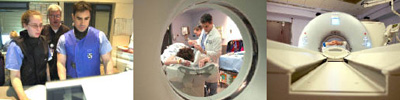
Cheryl Sadow, MD, technologist Richard Prior and Anthony Caravello, MD, operate Siemens Sensation Open CT at left and center; with BWH’s new Siemens Definition, at right.
Brigham and Women’s Hospital is not waiting until the Carl J. and Ruth Shapiro Cardiovascular Center opens next spring to make the latest imaging technology available to its patients.
In recent months, the hospital installed in CT L1 suite a Siemens Sensation Open, an interventional CT scanner that features an 82-centimeter opening and real-time imaging package software. In addition, CT L1 is home to the Siemens Definition, which uses Dual Energy technology or two imaging tubes to collect twice the amount of data as traditional CT machines. And in February, BWH installed a 64-slice CT scanner from Toshiba on the Nesson Pike.
“With tremendous commitment and support from our vendors and a dynamic and collaborative process that utilized the expertise of BWH’s physicians, nurses, technologists and administrators, we were able to secure top-of-the-line imaging equipment now while securing our ability to bring the best and most advanced technology of tomorrow to BWH,” Steven E. Seltzer, MD, chair of Radiology, said.
In addition to the imaging equipment recently installed at BWH, the Shapiro Center plans to feature a 256-slice CT scanner from Toshiba (once FDA approved), a second Siemens Definition with its Dual Energy technology, a 3.0 Tesla MRI made by Siemens, and a Discovery VCT PET/CT 64-slice scanner made by GE. All of these machines will be configured for cardiovascular imaging, offering BWH patients and clinicians an unprecedented array of imaging options under one roof.
According to Frank J. Rybicki, MD, PhD, co-director of Cardiovascular Imaging in the Department of Radiology, manufacturers and health care organizations are looking at two diverging technologies for the future of CT imaging. With its dual energy technology, Siemens is pushing for faster imaging, and Toshiba is increasing the volume of its image coverage by adding additional “slices” to its detector array.
“There are only a few hospitals with the Siemens Definition and only one other hospital moving forward with Toshiba’s 256-slice CT,” Rybicki said. “We will lead the way in cardiovascular care by having these imaging modalities together under one roof. Our patients will have access to a full array of imaging diagnostics.”
The Siemens Sensation Open and the Siemens Dual Energy in CT L1 in the last few months have enhanced patient care and imaging at BWH.
“These scanners represent the latest in CT technology, enhancing both our diagnostic imaging and interventional services,” said Stuart G. Silverman, M.D, director of Abdominal Imaging and Intervention, CT Scan and Cross-sectional Interventional Service.
CT L1: Siemens Sensation Open
An additional 12 centimeters—less than 5 inches—may not seem like a lot of extra space to the average person, but it represents a significant increase for a radiologist maneuvering catheters during an interventional procedure. The Siemens Sensation Open scanner features a larger bore or opening that leaves more room for such procedures around larger patients.
This scanner also allows for real-time imaging, so-called CT fluoroscopy, that helps interventional radiologists guide needles, catheters and therapy devices into the body. The CT scanner displays images of the abnormality to the radiologist in the room; devices are tracked using CT fluoroscopy. The scanner is used by the Cross-sectional Interventional Service to perform tumor and tissue biopsies, catheter drainage of fluid collections, and percutaneous tumor ablations.
“This scanner is the best interventional CT scanner that I have worked with to date,” Silverman said. “We’ve been performing CT-guided interventions for several years; this scanner helps us perform the procedures more effectively and efficiently.”
CT L1: Siemens Definition
BWH is the only Harvard-affiliated hospital equipped with the Definition. “The benefit to our patients is the ability to image non-invasively the coronary arteries, a large potential benefit over traditional coronary angiography,” said Rybicki. “The challenge for CT is to obtain speeds that rival projection images in conventional imaging, and the Definition has brought BWH far closer to that goal.” Patients can now be scheduled for noninvasive coronary imaging with this technology. The fact that there are two x-ray tubes and detector units simultaneously acquiring data gives BWH imagers a distinct advantage in imaging the coronary arteries. These advantages are expected to expand the clinical service and contribute to the changing role of coronary artery diagnoses.
“This represents a significant enhancement of our diagnostic imaging capabilities and a tremendous advancement in the care of our patients,” Rybicki said.
Imaging at Shapiro
Toshiba’s 256-slice CT scanner, which looks like a patient table surrounded by a massive, doughnut-shaped metal ring or central gantry, can scan four times the area scanned with a 64-slice CT. Its scanning area is 12.8 centimeters or five inches, a wide area that can capture the entire heart in one rotation.
“The gantry is so big, we literally will be scanning the entire heart in a heart beat,” Rybicki said. The 256-slice scanner will improve imaging for patients with arrhythmia, or irregular heartbeats, as well as studies of calcium buildup, artery hardening and blood flow.
Shapiro will house a combination of PET and CT technology in one machine, as well. GE has combined Positron Emission Tomography (PET) with Computed Tomography (CT) to enable detailed and motion-free images of the heart. By combining both modalities into one step, the system is especially suitable for cardiovascular applications.
“The integrated imaging of both the coronary arteries and flow of blood through the coronary arteries in the same setting is exceptionally powerful, as it facilitates not only diagnosis (like CT alone) but also management decisions by defining the potential need for revascularization,” said Marcelo Di Carli, MD, chief of Nuclear Medicine/PET.
BWH also will boast a powerful Siemens Trio 3.0 Tesla MRI dedicated to cardiovascular imaging in the Shapiro Center which will allow non-invasive study of functional information about coronary circulation. “This will give us exquisite vascular detail,” said Raymond Kwong, MD, of BWH’s Cardiovascular Division.