Researchers Develop New Tool to Help Brain Surgeons in Operating Room
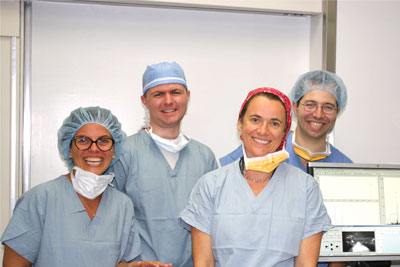
From left, Nathalie
Agar, Isaiah Norton, Alexandra Golby and Sandro Santagata comprise the BWH DESI
mass spectrometry team. |
Distinguishing the border between normal and cancerous brain areas can often
be difficult for surgeons attempting to remove tumors. But a new tool may change
this, allowing more comprehensive testing of brain tissue during surgery to help
surgeons quickly make the call between healthy and cancerous tissue.
So far, the tool has successfully identified the cancer type, severity and
tumor margins from tissue samples of five brain surgery patients, according to a
new study conducted by researchers from BWH’s departments of Neurosurgery,
Radiology and Pathology, and Purdue University.
“Tumor tissue within the brain often closely resembles normal brain tissue
and may have indistinct boundaries, so it is difficult to determine where tumors
end and normal brain tissue begins,” said co-study author Alexandra Golby, MD,
director of Image-Guided Neurosurgery in BWH’s Department of Neurosurgery, and
clinical co-director of BWH’s Advanced Multi-Modality Image Guided Operating
(AMIGO) suite.
“During surgery, we want to preserve as much functional brain tissue as
possible, especially when a tumor is in a critical area of the brain, such as
those that support movement, speaking or vision.”
Today’s surgical methods rely on a surgeon’s trained eye along with the help
of an operating microscope and brain image scans taken before or during surgery.
According to co-study author Sandro Santagata, MD, PhD, of BWH’s Department
of Pathology, examining frozen brain tissue specimens (which are transferred
from the operating room to a pathology laboratory) takes about half an hour—a
long time to wait during surgery. This problem is magnified when surgeons need
information from multiple samples during the course of a procedure.
A Tool in the Works
Attempting to develop a tool that would allow surgeons to address the time
delay that occurs during tissue analysis, researchers tested an imaging tool
known as “desorption electrospray ionization” (DESI) mass spectrometry.
DESI mass spectrometry was initially developed by researchers at Purdue
University. The BWH research team initiated a collaboration with Purdue
University to use this technology to test brain tissue samples from patients who
underwent surgery in the AMIGO suite and in standard BWH operating rooms.
In the study, surgeons removed 32 specimens from patients during surgery,
which were later analyzed by both the new tool and standard pathology methods to
test for accuracy. The researchers used DESI mass spectrometry to evaluate the
distribution and amounts of fatty substances, called lipids, within the brain
tissue specimens. A software program developed by the team then used the results
to characterize the brain tumors and detect boundaries between healthy and
cancerous tissue.
“The new tool is able, in a matter of seconds, to identify and classify many
types of brain tumors, and to recognize tumors that are likely to behave
aggressively,” said Santagata. “Accessing this type of information at a pace
that is more compatible with the pace of surgery could be a big plus for patient
care.”
Next Steps
The researchers plan to improve the classification software. BWH has set up
DESI mass spectrometry technology in the AMIGO suite and plans to test it to
detect brain and breast cancer margins during surgery.
“This approach could lead to real-time, image-guided surgery,” said Nathalie
Agar, PhD, director of the Surgical Molecular Imaging Laboratory in BWH’s
Departments of Neurosurgery and Radiology, and co-lead study author. “Such
extensive and detailed information about brain tissue that could lead to more
precise tumor removal was previously unavailable to surgeons. In addition,
having access to a detailed diagnosis on the day of surgery could help an
oncologist more efficiently design the course of post-surgery therapy.”

A new imaging tool has
successfully identified the cancer type, severity and tumor margins in patients
who underwent brain surgery in BWH’s AMIGO suite (pictured above). |